
Chromosomes, Genes, and DNA Worksheet Distance Learning Teaching
Chromosome number. Different species have different numbers of chromosomes. For example, humans are diploid (2n) and have 46 chromosomes in their normal body cells. These 46 chromosomes are organized into 23 pairs: 22 pairs of autosomes and 1 pair of sex chromosomes. The sex cells of a human are haploid (n), containing only one homologous.

Chromosome Structure
Chromosome - Definition, Structure, Function, Examples. Chromosomes are thread-like structures present in the nucleus. They are important because they contain the basic genetic material DNA. These are present inside the nucleus of plants as well as animal cells. Chromosomes were first discovered by Strasburger in 1815 and the term.

Provide me a diagram of chromosomes and genes. Brainly.in
Below the image is the label gametes made and there are 4 different gametes shown. One gamete is labeled uppercase a uppercase B, 25 percent and has one blue line with an uppercase A at the top and a uppercase B at the bottom of the line.. Homologous chromosomes are paired chromosomes that carry the same genes, but may have different alleles.
31 Label The Parts Of The Chromosome Labels Design Ideas 2020
What is a chromosome? Chromosomes are thread-like structures located inside the nucleus of animal and plant cells. Each chromosome is made of protein and a single molecule of deoxyribonucleic acid (DNA). Passed from parents to offspring, DNA contains the specific instructions that make each type of living creature unique.

Parts of Chromosome Diagram Quizlet
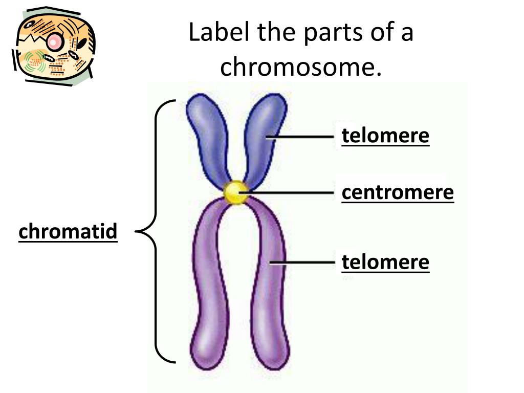
June 3, 2019 in Cell Biology, Genetics, Worksheets by Shannan Muskopf centromere, chromatid, chromosome, DNA, label, nucleus, practice, structure A diagram of a chromosomein the nucleus of the cell. Students label the chromatid, centromere, chromosomes, cell membrane, DNA, and nucleus.

Labeled Chromosome Structure Diagram imgprobe
The first number or letter used to describe a gene's location represents the chromosome. Chromosomes 1 through 22 (the autosomes) are designated by their chromosome number. The sex chromosomes are designated by X or Y. The arm of the chromosome.

Chromosome labeling with EdU. a Labeled region localization and
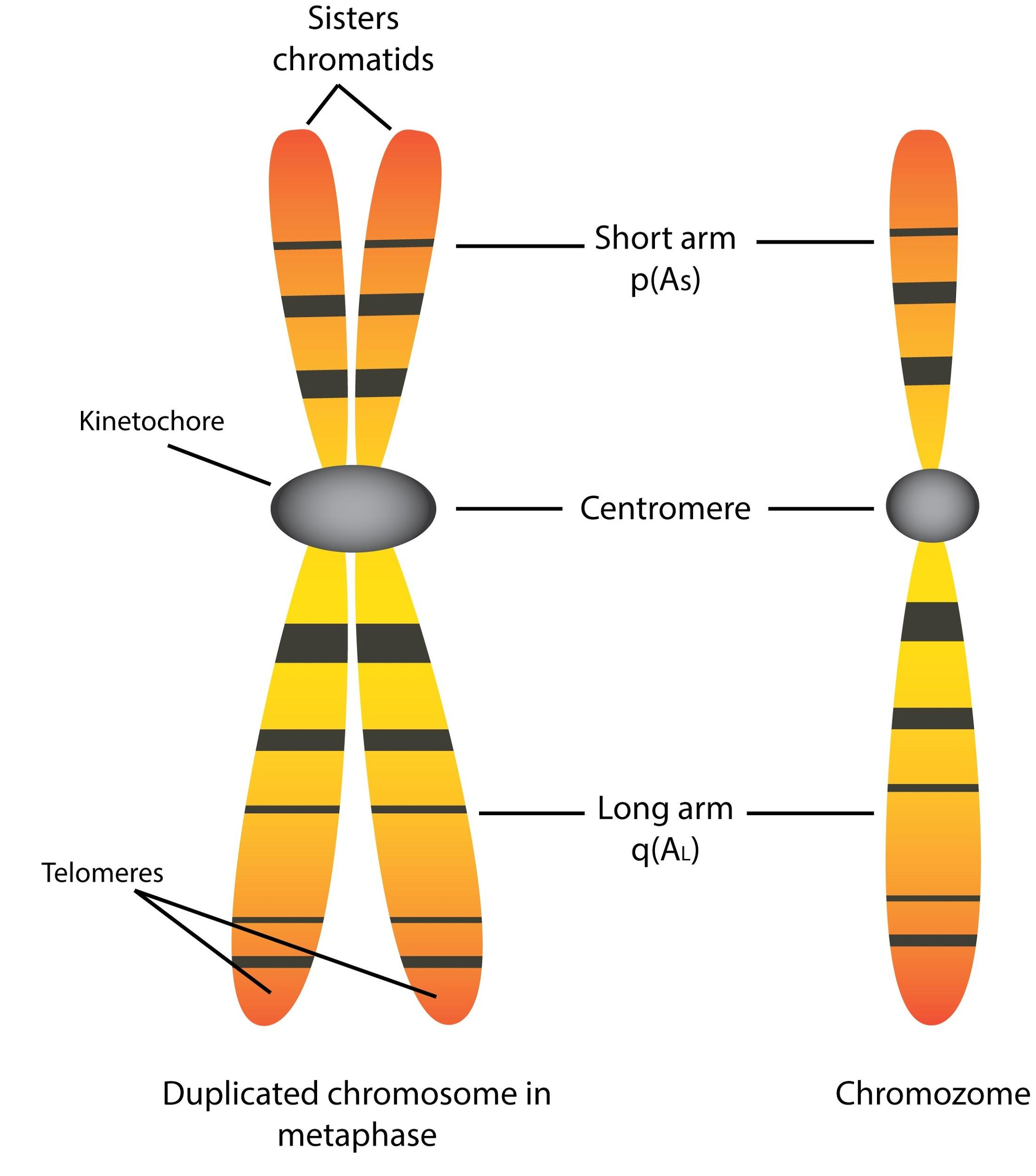
A karyotype is the number and appearance of chromosomes. To obtain a view of an individual's karyotype, cytologists photograph the chromosomes and then cut and paste each chromosome into a chart, or karyogram, also known as an ideogram. In a given species, chromosomes can be identified by their number, size, centromere position, and banding.

Chromosomes Definition, Structure, Functions & Example
The process of differentiating between cells is expressed by labeling the developmental tree. Tracing a path from the root to a specific cell in the tree reveals the history of its divisions. Normal human fetal cells will divide approximately 40 to 60 times before cell division halts as demonstrated by Hayflick [ 1 ].

Mitosis Worksheet And Diagram Identification Answers
Students label a simple diagram of a chromosome showing the centromere, chromatid, DNA, and the location of the chromosome within the nucleus of a cell.
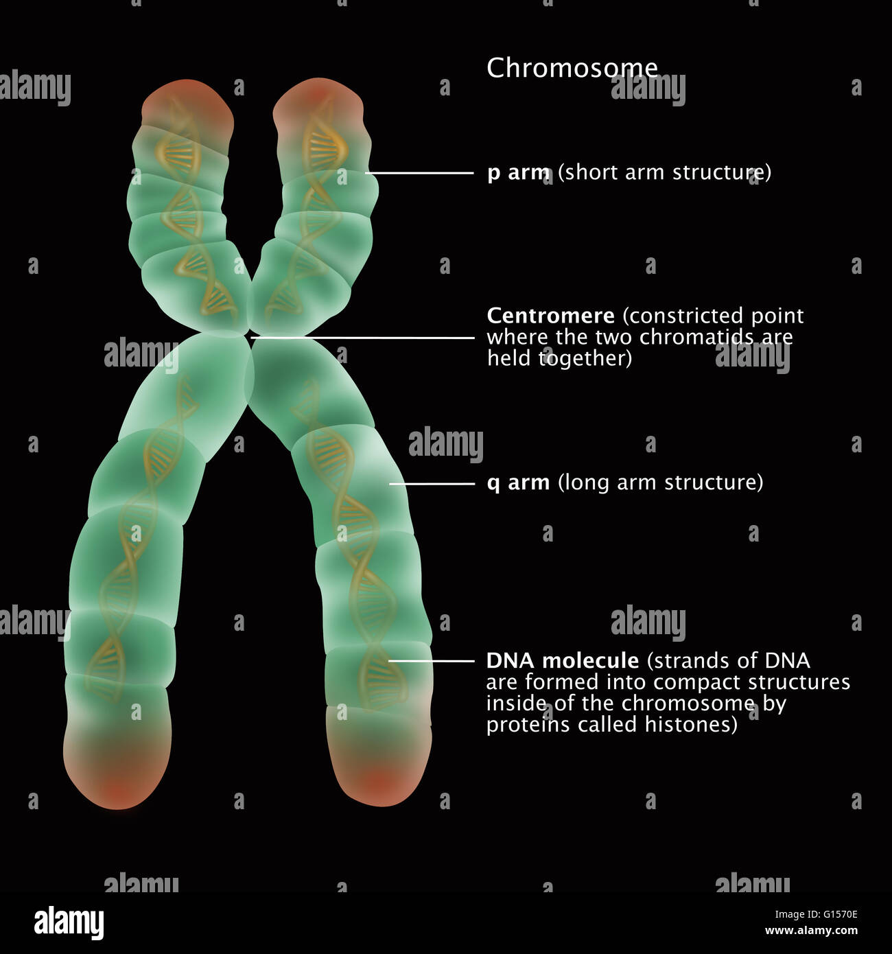
Illustration of the detailed structure of a chromosome. The p arm
Since all of the cells in an organism (with a few exceptions) contain the same DNA, you can also say that an organism has its own genome, and since the members of a species typically have similar genomes, you can also describe the genome of a species.

Locus the location of a gene on a chromosome or on a linkage map
In this paper, we describe the labeling of human genomic loci in live cells with three orthogonal CRISPR/Cas9 components, allowing multicolor detection of genomic loci with high spatial resolution, which provides an avenue for barcoding elements of the human genome in the living state.
Chromatid is(a) One half of chromosome(b) Haploid chromosome(c
Mitosis consists of four basic phases: prophase, metaphase, anaphase, and telophase. Some textbooks list five, breaking prophase into an early phase (called prophase) and a late phase (called prometaphase). These phases occur in strict sequential order, and cytokinesis - the process of dividing the cell contents to make two new cells - starts.

Chromosome structure Chromosome, Chromosome structure, Structural biology
A chromosome is a thick ribbon-like structure containing the genetic material i.e., DNA made up of genes. This information is necessary for maintaining and making more copies of a cell.

Basic components of chromosome Telomeres, Cell Division, Chromosome
The process of differentiating between cells is expressed by labeling the developmental tree. Tracing a path from the root to a specific cell in the tree reveals the history of its divisions. Normal human fetal cells will divide approximately 40 to 60 times before cell division halts as demonstrated by Hayflick [ 1 ].
For Keeping X Chromosomes Active, Chromosome 19 Marks The Spot 04/17/2017
ADVERTISEMENTS: The following points highlight the six main parts of a chromosome. The parts are: 1. Pellicle and Matrix 2. Chromatids, Chromonema and Chromomeres 3. Centromeres 4. Secondary Constriction 5. Satellite 6. Telomere. Part # 1. Pellicle and Matrix: A membrane which surrounds each chromosome is said as pellicle. A jelly substance present inside the […]

Chromosome labeling with EdU. a Labeled region localization and
Although currently CRISPR technology is mostly applied to gene editing and regulation 1, we and others have used it to label defined chromosomal loci to image the three-dimensional structure of.
