
Rat Dissection And Anatomy at Arizona State University West Campus StudyBlue
Label the diagram using the descriptions and bold words. Trace the flow of blood using arrows. Branches of the Aorta The aorta has four general areas. Locate each of these on your rat. ascending aorta - the upper part of the vessel that starts at the atrium aortic arch - the place where the aorta bends to the left.
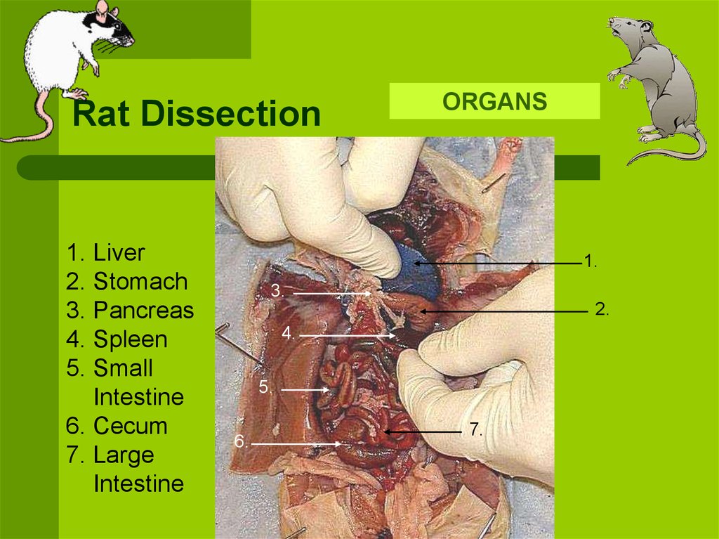
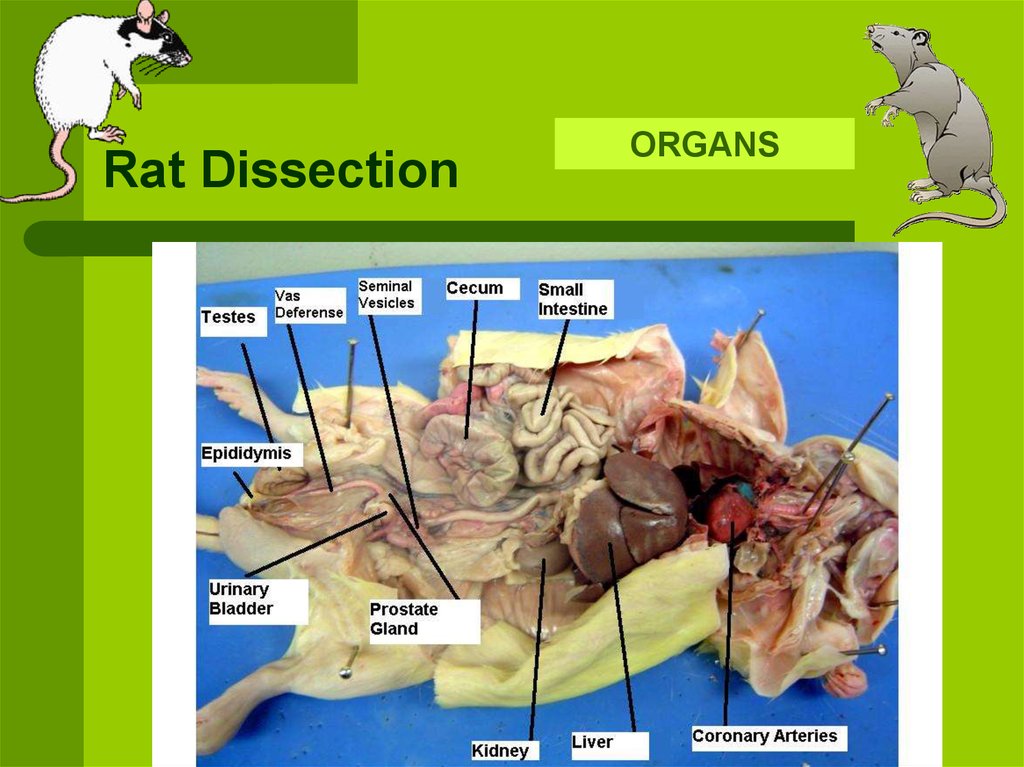
Rat Dissection Online Presentation 984
Phalanges Skeletal System Cervical vertebrae Thoracic vertebrae Lumbar vertebrae Sacrum Illium Ischium Caudal vertebrae Pubis Femur Rib Patella Tibia Fibula

Rat (Brown) Exploring Nature Educational Resource
Tap or move your mouse over the rat diagram below to view the organ names. *Tip: on smaller devices, you may want to flip your device to landscape if you can't see the full width of the image. Medical Illustration by Chris McKee for the Rat Guide. Additional descriptions will be added by the Rat Guide Team.

Investigation Rat Dissection Biology LibreTexts
The Anatomy and Physiology of Laboratory Rat Saurabh Chawla & Sarita Jena Chapter First Online: 24 July 2021 2776 Accesses Abstract The laboratory rat is commonly used as an experimental model in biomedical research.
Anatomy Of The Rat Anatomical Charts & Posters
In this video, I talk you through how to do a dissection of a rat to highlight the major mammalian organ systems. It shows the excretory system, digestive sy.
Rat Anatomy by izzycreates on DeviantArt
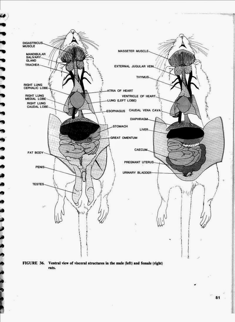
1. Locate the diaphragm, which is a thin layer of muscle that separates the thoracic cavity from the abdominal cavity. 2. The heart is centrally located in the thoracic cavity. The two dark colored chambers at the top are the atria (single: atrium), and the bottom chambers are the ventricles.

Rat dissection labeling (pt 1) Diagram Quizlet
Rats are often used in dissection classes because they are readily available and they possess the typical mammalian body plan. Most of what you learn on the rat is applicable to the anatomy of other mammals, such as humans.. The diagram below illustrates the muscles of the ventral surface of the rat. Be able to identify those
PPT Rat Dissection PowerPoint Presentation, free download ID638753
The wedge-shaped area on the central portion of each lung where the bronchi, arteries, veins, and nerves enter and exit the lungs. Lines abdominopelvic cavity. Anatomy rat dissection quiz- labeling parts Learn with flashcards, games, and more — for free.
Rat Dissection Anatomy Anatomical Charts & Posters
Availability: Rats are most frequently used as type specimen for mammalian dissection because they are readily available and they possess the typical mammalian body plan. Most of what you learn on the rat is applicable to the anatomy of other mammals, such as humans. Rats can be obtained from pet shops, biological supply companies or pharmaceutical firms.
Rat Dissection Biology 11 Honours Animalia Labs
Rat Navigation Step 1: Body Regions Step 2: External Features Step 3: Expose the Muscles Step 4: Expose the Bones Step 5: Head & Neck Step 6: Thoracic & Abdomen Step 7: Urogenital System Student handouts for rat dissections: This is a walk-through of the rat dissection with photos showing the key features of the rat.
[DIAGRAM] Female Rat Reproductive System Diagram
Dissection of Genital System. The rat is a typical mammal. Formerly guinea pig (Cavia sp.) (Fig. 19.1) were used for dissection in most of the undergraduate and postgraduate colleges in Indian Universities. Of late, due to prevailing high cost, guinea pig is being replaced by rat. Four species of rats are common in India, of which three are wild.
Rat Dissection online presentation
61 Share 7.9K views 2 years ago #howtodraw #rat #rat #ratdiagram #howtodraw Students need to learn about the basic parts of a rat. So in this video, I try to help you with drawing a labeled.

Rat External Anatomy Diagram Quizlet
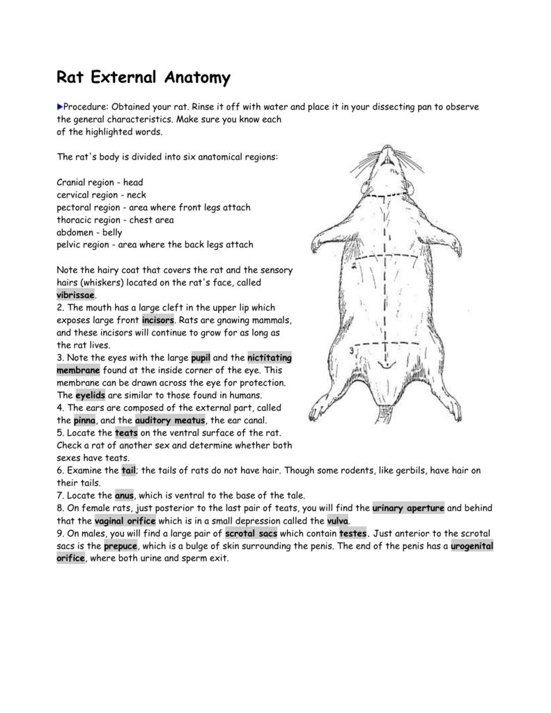
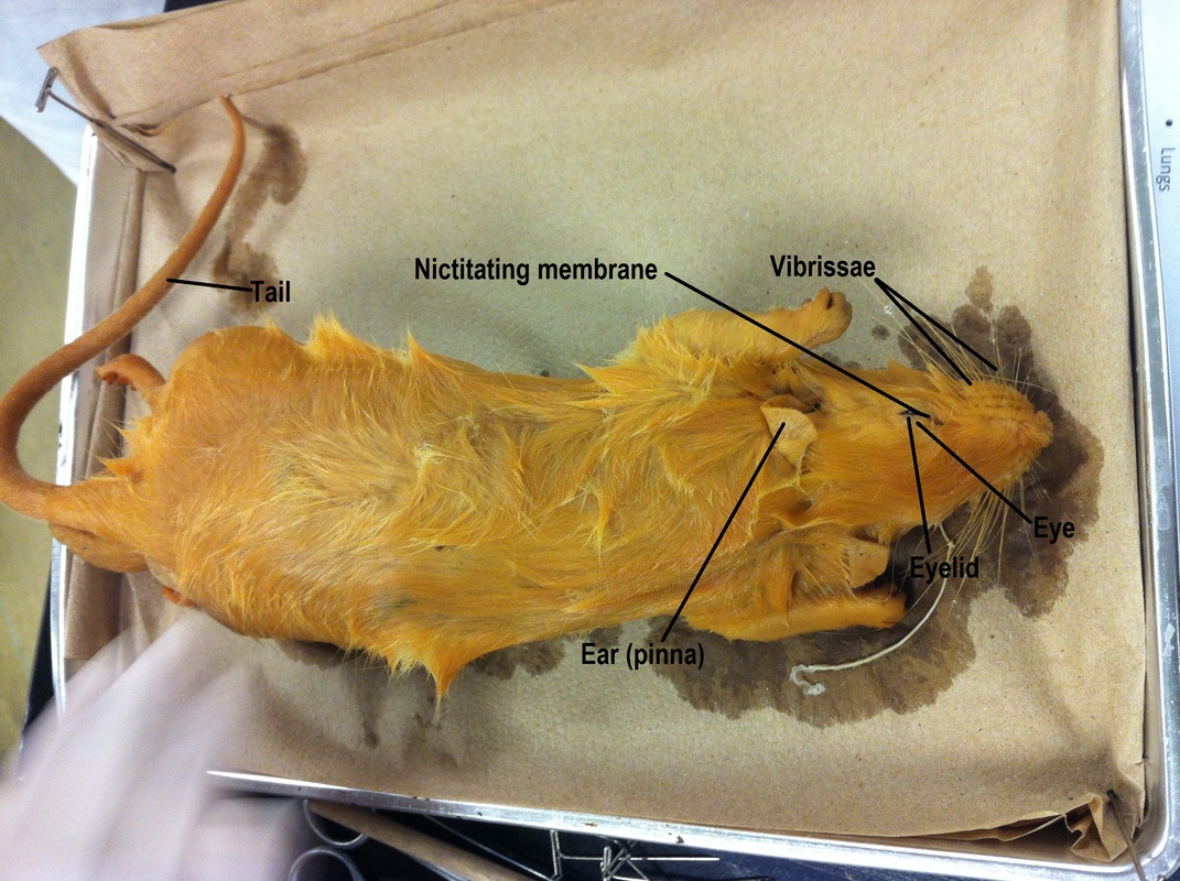
The external anatomy encompasses the head (cranial region), neck (cervical region), chest (thoracic portion), pectoral (portion where forelegs attach), abdomen, pelvic (portion where hind legs attach), and tail. Though both sexes have teats, you can differentiate the male rat from the female rat.

The rat's muscle anatomy. The rat's muscle anatomy is shown in... Download Scientific Diagram
Locate the delicate ureters that attach to the kidney and lead to the bladder. Wiggle the kidneys to help locate these tiny tubes. Procedure: Remove a single kidney (without damaging the other organs) and dissect it by cutting it longitudinally. Locate the cortex (the outer area) and the medulla (the inner area).
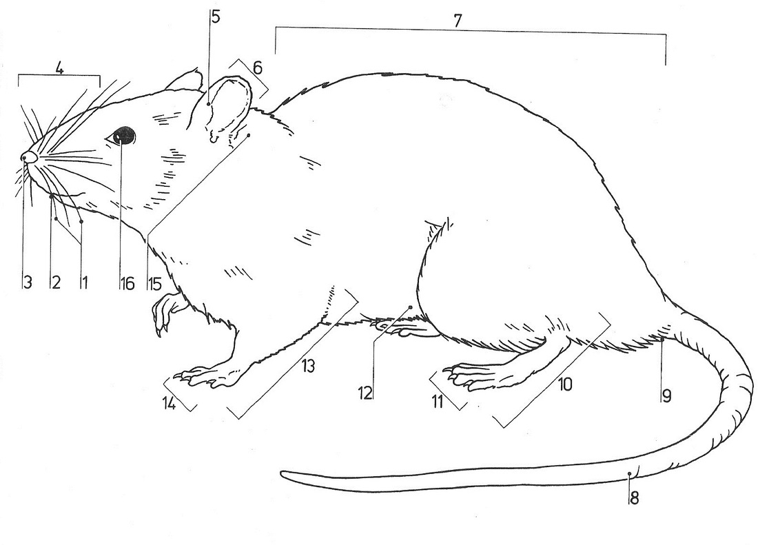
Proper rat anatomy is very important. Can you identify these parts on yours? RATS
Female Reproductive System. The organs of the female reproductive system are specialized to produce ova (eggs), transport the egg cells to the site of fertilization, to provide a favorable environment for developing embryos, and to move offspring outside of the body (birth) at the appropriate time. The reproductive system also supplies.

Rat Anatomy Poster Bones Organs Small Mammal
Conceptually, a neural circuit, network, or system may be viewed as a set of nodes or "parts"—including gray matter regions, neuron types, individual neurons, and synapses, considered at descending, nested levels of granularity—with a set of connections between nodes.
