
Frog Heart Diagram Quizlet
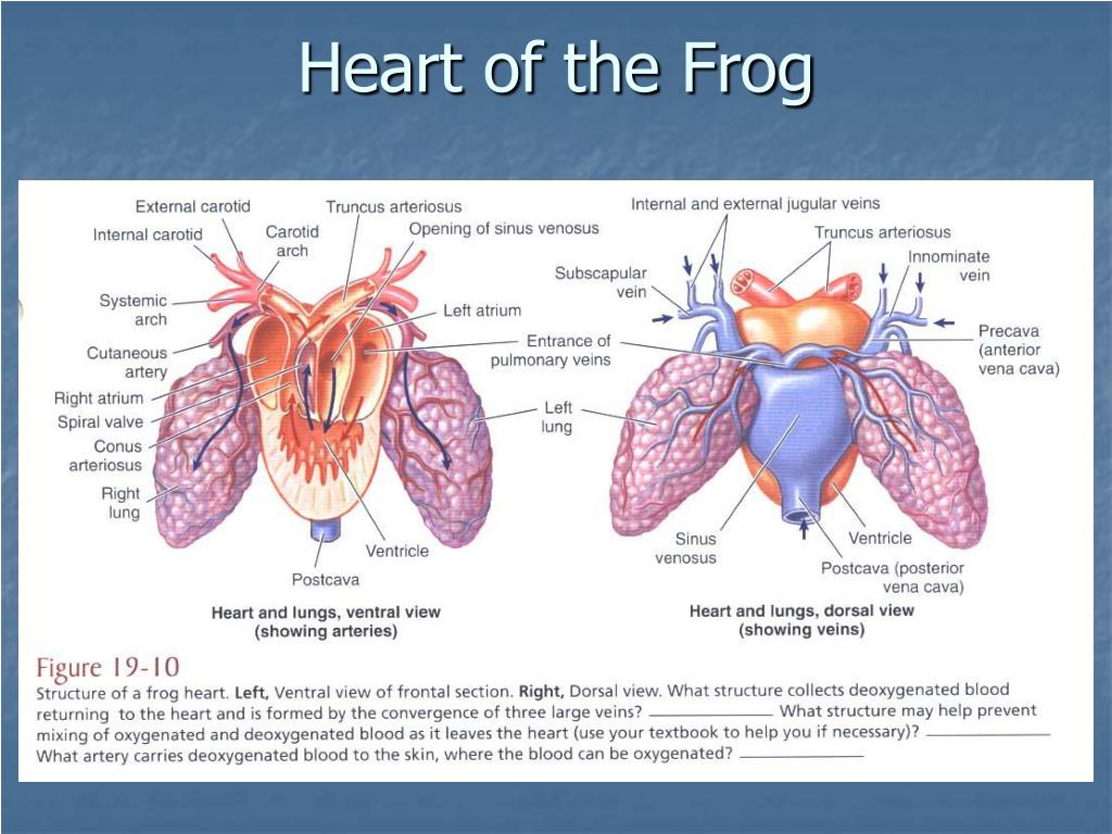
I. Introduction. The heart of the frog has three chambers, one ventricle and two atria. Blood leaves the heart from the ventricle through a single truncus arteriosus which is short and soon branches into two aortic arches which loop left and right and dorsal to the heart to rejoin as a single aorta in the mid dorsal region of the body cavity.

Important images for system of frog Agrivetforestry Entrance
Circulatory system - Amphibians, Blood Vessels, Heart: Modern amphibians are characterized by the flexibility of their gaseous exchange mechanisms. Amphibian skin is moistened by mucous secretions and is well supplied with blood vessels. It is used for respiration to varying degrees. When lungs are present, carbon dioxide may pass out of the body across the skin, but in some salamanders there.

Illustrations and Movies iWorx Systems Inc
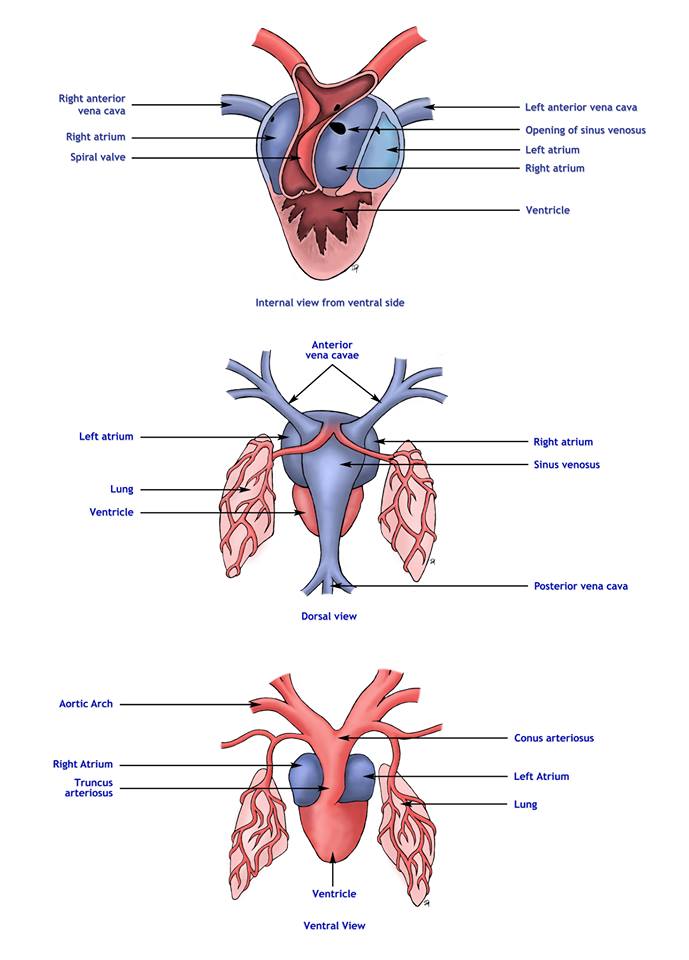
Frogs are amphibians and have a closed circulatory system. Unless there is an abnormal mutation present, frogs only have one heart to pump blood throughout the body. A frog has a three-chambered heart. The chambers include two atria and a ventricle. The right atrium receives deoxygenated blood from the veins.

Standard Note Structure and working of Frog's heart
The heart of the African clawed frog has a double-inlet and single-outlet ventricle supporting systemic and pulmonary circulations via a truncus, and a lifespan of 25-30 years. We sought to understand the unique cardiac anatomic and physiologic characteristics, with balanced circulation and low metabolic rate, by comparing the basic anatomy.
Frog Heart ClipArt ETC
Frogs are a type of amphibians with a closed circulatory system.Hence, its blood only circulates through the blood vessels and heart. The circulatory system of frogs composes of two parts: the cardiovascular system and the lymphatic system.The main function of the cardiovascular system is to supply oxygen and nutrients to the tissues and to aid in the elimination of metabolic wastes while the.
PPT Vertebrata PowerPoint Presentation, free download ID65868
A frog's digestive system starts with their long, sticky tongues that they use to catch their prey. Inside their mouth, frogs also have small teeth, and a set of two larger teeth. These are not really used to chew since frogs swallow their prey alive and whole. Their teeth are used mostly to keep back their prey.
PPT Frog Body Parts and Functions PowerPoint Presentation ID1266919
The left auricle then contracts, sending the oxygenated blood into the ventricle, which in turn contracts and sends the blood through the bulbus arteriosis into the arteries of the body. Anatomical model of a dissected frog: 1 Right atrium, 2 Lungs, 3 Aorta, 4 Egg mass, 5 Colon, 6 Left atrium, 7 Ventricle, 8 Stomach, 9 Liver, 10 Gallbladder, 11.

Class 11Zoology LecturesDiscuss the structure of the frog's heart
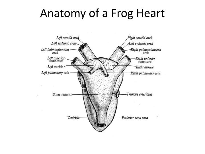
Detailed Structure of Frog's Heart. Heart of frog is three chambered. It is dark red colored conical muscular organ situated mid-ventrally in the anterior part of the body cavity in between two lungs. The heart is enclosed in two membranes- an inner epicardium and outer pericardium. The space between these two layers is called pericardial cavity.
Frog Heart Diagram Back View
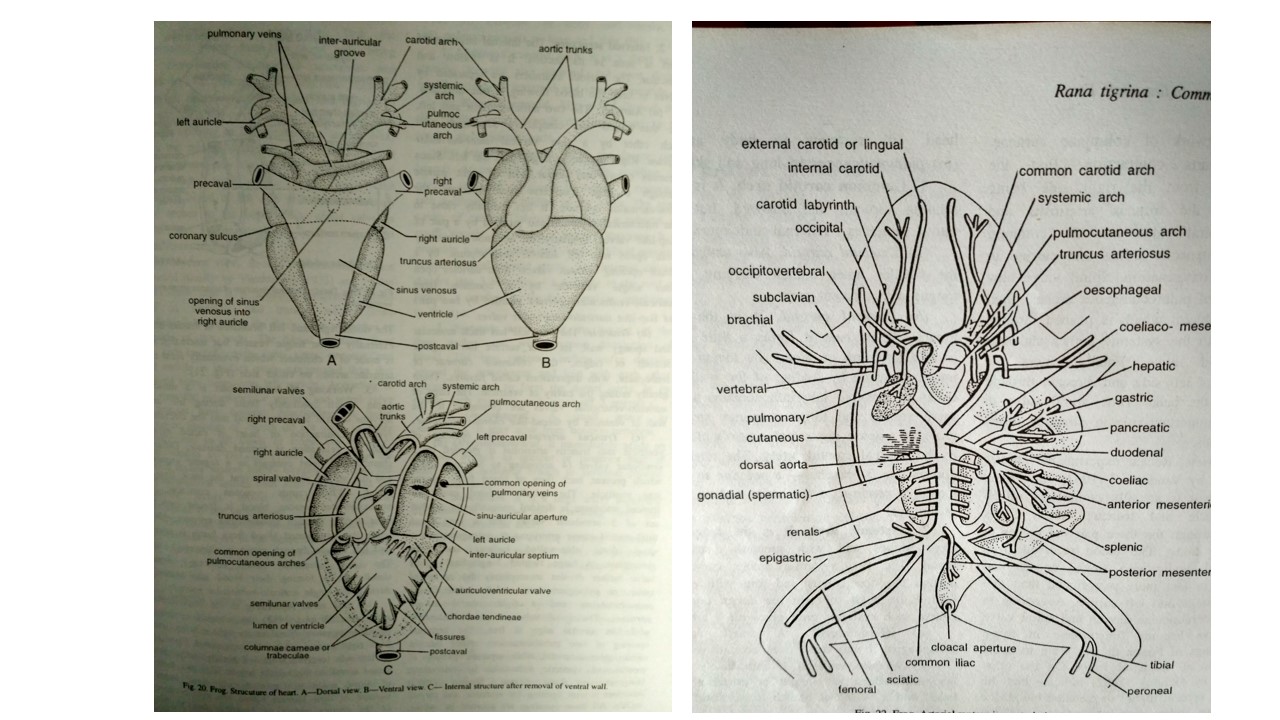
The blood vessels which carry the blood away from the heart to different parts of the body constitute the arterial system. In frog arterial system begins with the truncus arteriosus which divides into two large right and left branches or trunks. Each branch divides into three aortic arches- an anterior carotid arch, a middle systemic arch and a posterior pulmocutaneous arch. Carotid Arch: The.
.jpg)
Notes Guide Book Describe the ventral and dorsal side of the frog's heart.
Frog Artery. The primary function of the heart is to pump oxygen rich blood to organs such as the brain, liver, and kidneys as well as all other tissue. The heart of the frog is different from the hearts of warm-blood animals such as humans. Although mammals have four chambers, amphibians, which are cold-blood animals, have only three.
frogs circulatory system
This study guide is easy to understand, yet has thorough information including a downloadable diagram of a frog's circulatory system and heart. Also covered is a full description of how the frog's three-chambered heart works. If you need to learn about this topic for a school project in science or biology, or you are just interested in frogs and their anatomy, then you will value.

how to draw frog heart.....dorsal view YouTube
A diagram of a frogs heart. A look inside the heart of a Frog looking at key valves and parts of the heart.

how to draw frog heart.....ventral view YouTube
blood circulation in frog. The blood vascular or circulatory system of frog is closed. The circulatory system consists of: Heart: Arterial system. Venous system. Blood. Lymphatic system. Its main function is to transport all essential liquid and gaseous materials to the living tissues.

Frog Heart Anatomy Anatomy Book
The blood vascular system of frog is closed. It includes the heart, blood vessels, blood and lymphatic system. The prime function of this system is to distribute the digested food and oxygen to different parts of the body, in order to release energy to carry out life activities and also to bring the excretory and gaseous wastes to organs of.
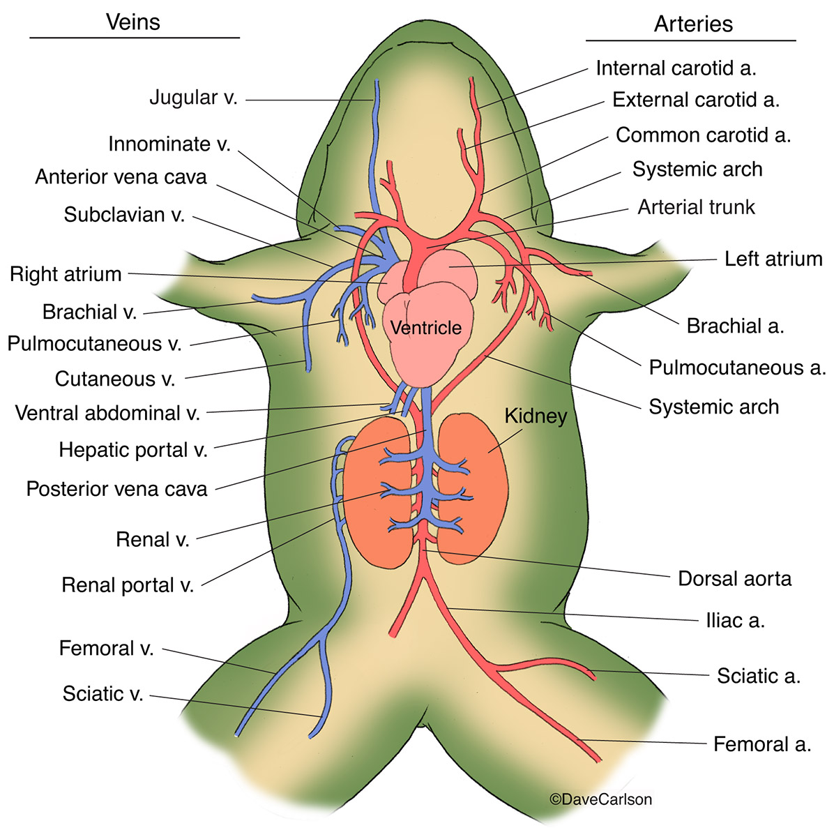
Frog Generalized Circulatory System Carlson Stock Art
Article Shared by. ADVERTISEMENTS: In this article we will discuss about the origin of the frog heart beat with the help of suitable diagram. By separating atria from the ventricles of excised hearts, William Harvey in 1628 had shown that the atrial rhythm was higher than the ventricular rhythm. Keith and Flack (1907) have described that from.
Heart of a Frog ClipArt ETC
The heart of the African clawed frog has a double-inlet and single-outlet ventricle supporting systemic and pulmonary circulations via a truncus, and a lifespan of 25-30 years. We sought to understand the unique cardiac anatomic and physiologic characteristics, with balanced circulation and low metabolic rate, by comparing the basic anatomy structures with focused echocardiography and.
